Ebstein anomaly
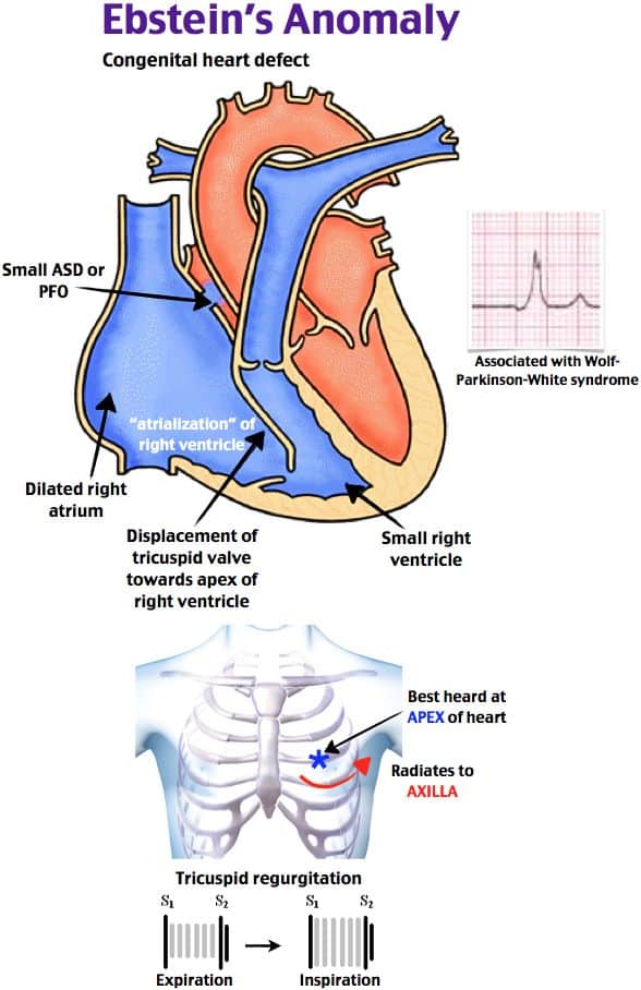
Ebstein anomaly is a rare congenital heart disease in which the tricuspid valve of the heart is placed far down into the right ventricle. This abnormal valve is prone to blood regurgitation into the right atrium. This may increase the pressure inside the right atrium and lead to progressive right atrial enlargement. Regular monitoring of the heart is indicated if this anomaly is present. The treatment for this anomaly is usually given if a patient starts to feel the symptoms of an enlarged heart.
Epidemiology of Ebstein anomaly
As it is a very rare congenital cardiac anomaly, the actual incidence rate is not very well-reported. Research revealed 1 in 0.2 million live births may have Ebstein anomaly. It accounts for <1% of all congenital heart anomalies. Both genders are equally affected. The incidence among the caucasian population is slightly higher than the other races.
Ebstein anomaly causes
The exact causes of the Ebstein anomaly are not fully understood. But research has revealed genetic factors, environmental factors, and positive family history is associated with Ebstein anomaly. The use of benzodiazepines and lithium during pregnancy is associated with Ebstein anomaly.
Signs and symptoms of Ebstein anomaly
The clinical features of Ebstein anomaly mostly develop due to heart enlargement and heart failure. These symptoms are –
- Shortness of breath because the failing heart cannot effectively circulate enough blood to the lungs and body.
- Cyanosis or blue discoloration of the skin when the percentage of carboxy-hemoglobin builds up.
- Ascites and leg edema as a result of right heart failure.
- Palpitations and arrhythmias.
- Fatigue
- Cough if pulmonary edema develops.
- Higher chance of stroke, bacterial endocarditis, and pulmonary embolism.

Associations with Ebstein anomaly
- Wolff-Parkinson White syndrome
- Atrial septal defect
- Patent foramen ovale
- William’s syndrome
Diagnosis of Ebstein anomaly:
Clinical examination
- Signs of heart failure – cyanosis, limb edema, pulmonary edema, S3 heart sound
- Signs of arrhythmia – irregular pulse
- Signs of tricuspid regurgitation – a systolic murmur, and systolic thrill.
- Signs of infective endocarditis – fever, splinter hemorrhage, Osler nodes, Janeway lesion, Roth spots, etc.
Investigation
- ECG of Ebstein anomaly can detect
- Abnormal heart rhythm can be present.
- P-pulmonale (tall p-wave) due to enlarged right atrium,
- Short PR interval, and delta wave may be present if WPW syndrome co-exists.
- Right axis deviation.
- ECG of Ebstein anomaly can detect
- Echocardiogram of Ebstein anomaly can detect
- Enlarged heart size
- Tricuspid regurgitation
- Presence of atrial septal defect and patent foramen ovale
- Ejection fraction
- Presence of any embolus or infective foci
- Echocardiogram of Ebstein anomaly can detect
- Chest X-ray can detect
- Pulmonary edema
- Enlarged heart shadow
- Kerley’s B lines
- Chest X-ray can detect
- MRI of the cardiac region
Differential diagnosis of this anomaly
- Tricuspid regurgitation.
- Atrial septal defect.
- Tricuspid atresia.
- Wolff-Parkinson -white syndrome.
Ebstein anomaly treatment
Proper monitoring
People with milder symptoms need no treatment, but you need proper follow-up with your doctor to control this. Regular ECG and Echocardiogram is needed to observe for any possible complications developing.
Diet
A heart-friendly diet is recommended. If heart failure develops then a fluid and salt restriction diet may be recommended.
Medicine/ surgery
Patients undergo lifetime cardiac monitoring after being diagnosed with Ebstein anomaly. The treatment is complex and often involves a multidisciplinary team. The treatment usually depends on the patient’s symptoms and development of complications. Some examples are given below –
- If the patient develops heart failure then heart failure medications like beta-blockers, ACE inhibitors, etc can be given.
- If patients develop arrhythmia then antiarrhythmics can be given. Any aberrant electrical pathway in the heart can be surgically ablated.
- If the patient develops infective endocarditis then antibiotics are prescribed. If antibiotics don’t work and the patient develops abscess then surgery may be done to remove the abscess.
- Blood thinners can be given if there is a history of blood clots.
- If tricuspid regurgitation is too severe then tricuspid valve replacement may have a role.


